
Figure 3. Vertex-wise analysis of CTh in ALS patients with higher UMN burden compared to HC.
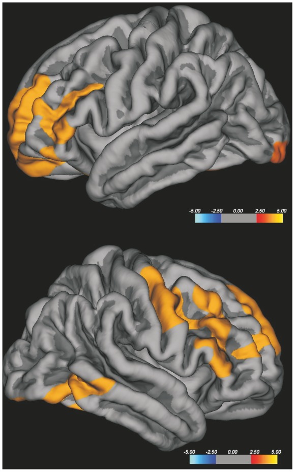
ALS patients with higher UMN burden showed cortical thinning in the right precentral gyrus, in bilateral superior, middle and inferior frontal gyri, in the right inferior temporal gyrus and in left lateral occipital cortex. The colour bar scale represents t values.