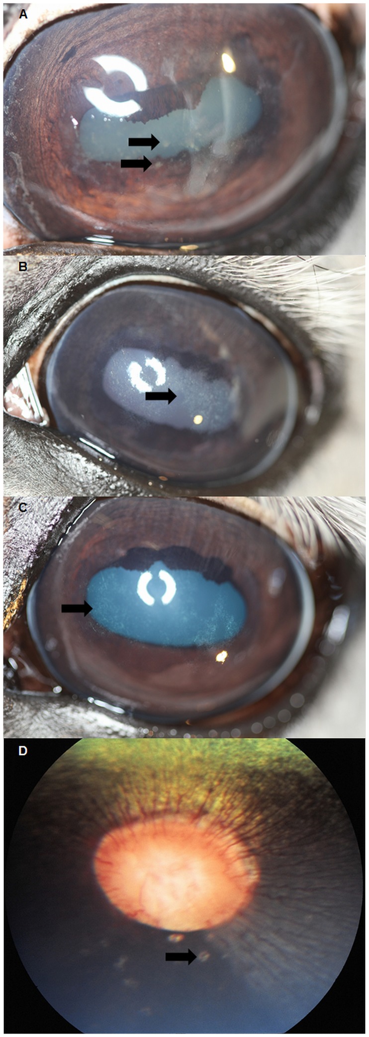
Figure 1. Ophthalmic findings of inflammatory origin.
A: Photograph of stallion diagnosed with superficial punctate keratitis with associated corneal oedema. Note the arrow pointing to superficial punctate infiltrates. B & C: Photograph of stallions diagnosed with anterior stromal opacities, suggestive of antecedent corneal inflammation. Note the arrows pointing towards the geographic areas of anterior stromal opacity. D: Fundus photograph of a stallion diagnosed with “bullet-hole” lesions in the peripapillary area. Note the arrows pointing towards the peripapillary “bullet-hole” lesions.

