
Figure 4. Circulating hemocytes and PCT cells are labeled for histamine.
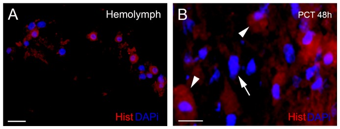
(A) Histamine-positive cells are red in the hemolymph; and the nuclei, DAPI stained, are in blue. (B) Injured PCT (48 h after eyestalk ablation) showing cells labeled for histamine (red) and DAPI (blue). The majority of histamine-positive cells contain granules (arrowheads) and round nuclei. No labeling was seen in the elongated nuclei (arrow) of the cells with morphology of glial cells. Scale bars: A = 20 µm; B = 10 µm.