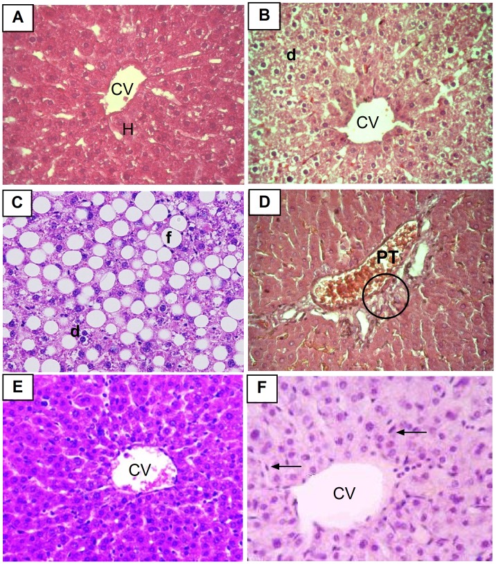
Figure 7. Photomicrographs of liver sections stained by H & E (×400).
A: liver section of normal rat shows normal central vein (CV) and surrounding hepatocytes (h). B: liver section of arthritic non-treated rat shows degeneration in hepatocytes (d). C and D: liver sections of arthritic rat treated with MTX alone shows severe fatty change (f), degeneration (d) of hepatocytes, congested portal tract (PT) and lost cell boundaries with distortion of normal architecture (circle). E: liver section of arthritic rat treated with BV alone shows normal hepatic architecture. F: liver section of arthritic rat concurrently treated with MTX and BV showing mild congestion in the central vein (CV) with diffuse kupffer cells proliferation (arrow) in between hepatocytes.

