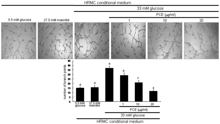
Figure 5. Suppression of endothelial tube formation by PCE.
HRMC were incubated in 5.5[with (w/) 27.5 mM mannitol and w/33 mM glucose] for 8 h in the absence and presence of 1–20 µg/ml PCE. Tube formation of HUVEC was assayed using matrigel. Cells were fixed and microphotographic images were captured at ×100 magnification. The branching points were continuously monitored and the number of tubes was counted from the images. Multiple five random fields of view were analyzed for the quantitative results (mean ± SEM, n = 4). Respective values not sharing a letter are different at P<0.05.

