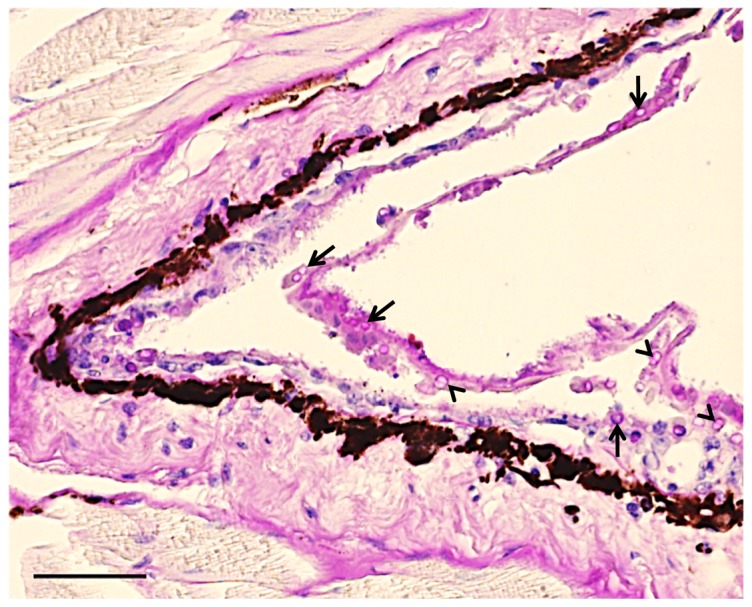
Figure 2. Skin histological section of a wild southern Darwin’s frog (Rhinoderma darwinii) with cutaneous chytridiomycosis.

Note multiple empty zoosporangia (arrows) within the superficial keratinised layer of the epidermis. Several zoosporangia with an internal septum can be seen (arrowheads), morphologically typical of Batrachochytrium dendrobatidis. Stained with Periodic Acid-Shiff (PAS). Bar = 20 µm.
