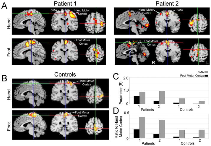
Fig. 1.
(A) fMRI activation maps of patients performing the hand motor task (top row) and the foot motor task (bottom row). Synkinesia patients had prominent activity in the supplementary motor area and foot motor cortex during the hand motor task. Patient 1 was right handed, and patient 2 was left handed; images are displayed in radiological convention. (B) Average fMRI activation maps of two controls performing the hand motor task (top row) and the foot motor task (bottom row). (C) Activation of mesial cortical regions of interest from the supplementary area (SMA) and the foot motor cortex during performance of the hand motor task. Patients with synkinesia both had greater activation in these areas compared to controls. (D) The ratio SMA or foot motor cortex activation to hand motor cortex activation during performance of the hand motor task. Patients with synkinesia had higher ratios of SMA and foot motor cortex activity than controls, suggesting that increases in activation could not be explained by increased cortical excitability.

