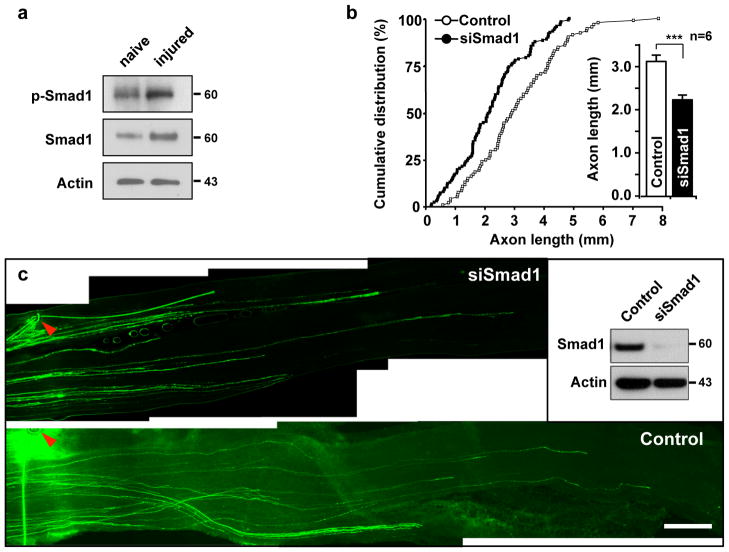
Figure 8. Acute depletion of Smad1 in vivo prevents sensory axon regeneration.
(a) Representative immunoblots of DRG lysates from naïve or injured mice. Mice were subjected to sciatic nerve transection to induce injury. DRGs were collected at 3 days after injury and subjected to Western blot analysis. Representative immunoblots for phospho-Smad1 (p-Smad1) and Smad1 are shown. Actin antibodies were used as a loading control.
(b, c) L4 and L5 DRGs of an adult mouse were electroporated in vivo with either EGFP alone (control) or together with siRNAs against Smad1 (siSmad1), as indicated, followed by sciatic nerve crush at 2 days after the electroporation. Axon regeneration was assessed by whole-mount analysis at 3 days after nerve injury. In vivo electroporation and investigation of axon regeneration were performed as depicted in Fig. 3a. Using whole-mount nerve segments, the lengths of all identifiable regenerating axons were measured from the crush site (marked by epineural suture, arrowheads) to the distal axon tips. For quantification, lengths of a total of 92 axons from six control mice and 121 axons from six siSmad1-transfected mice were measured. Quantification (b) and representative images (c) of regenerating axons are shown. The inset shows knocking down of Smad1 by siSmad1 in adult DRG neurons in vivo. Scale bar, 500 μm. Error bars represent s.e.m. * p < 0.05, unpaired two-tailed student t test. Original immunoblot images are shown in Supplementary Fig. S12.

