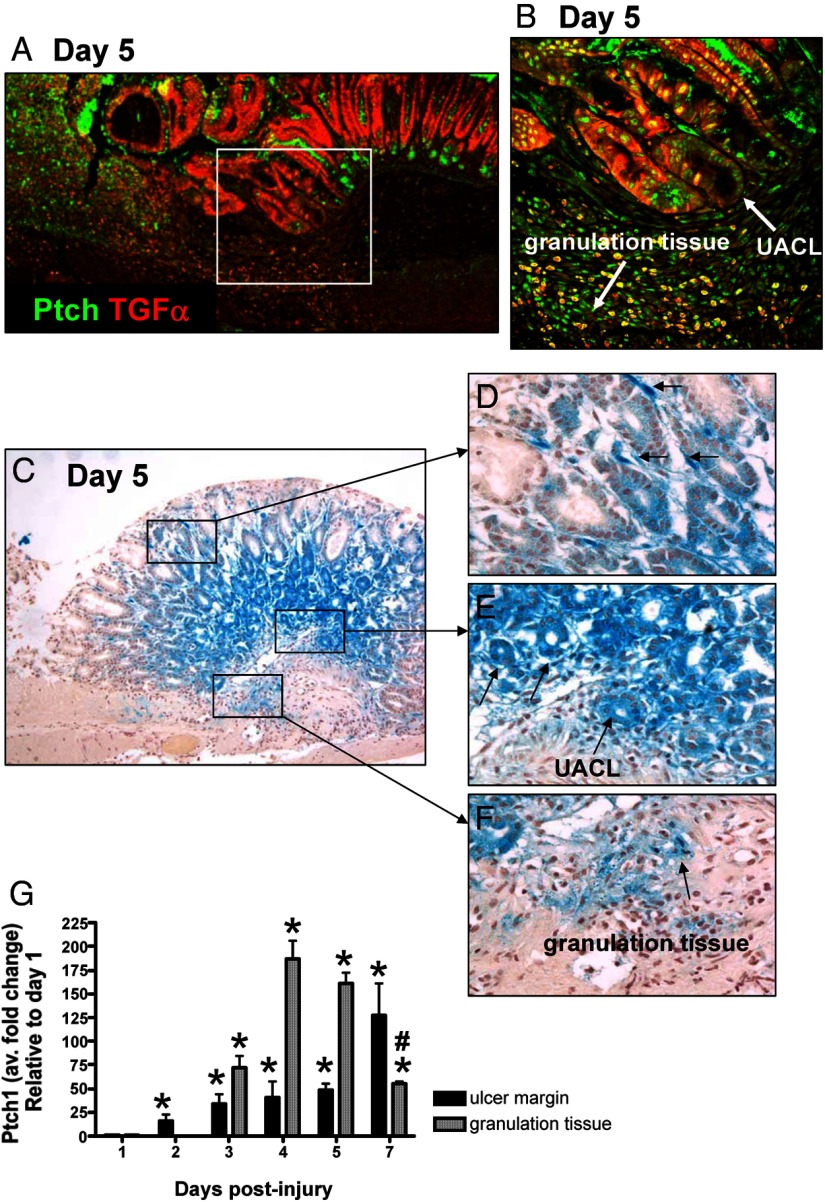
Figure 4.
Expression of Ptch within the healing zone of the injured stomach. A, Immunofluorescence staining using an anti-Ptch and anti-TGFα antibodies of stomach sections collected from a control mouse 5 days after acetic acid-induced injury. Ptch expression (green) colocalized with TGFα-positive (red) UACL at the ulcer margin and within the granulation tissue. Higher magnification is shown in B. C, β-Galactosidase activity in the gastric mucosa of control mice 5 days after the acetic acid-induce injury using the heterozygote B61(29-Ptch1tm1Mps/J Ptch reporter mice). X-gal staining demonstrating Ptch1 expression shown within the mesenchyme (D), at the ulcer margin (E) and within the granulation tissue (F). G, qRT-PCR was performed using RNA collected from the ulcer margin and granulation tissue using LCM. Data are expressed as the mean ± SEM relative to day 1 with three to four mice per time point. *, P < .05 compared with day 1; #, P < .05 compared with days 4 and 5.

