Figure 1.
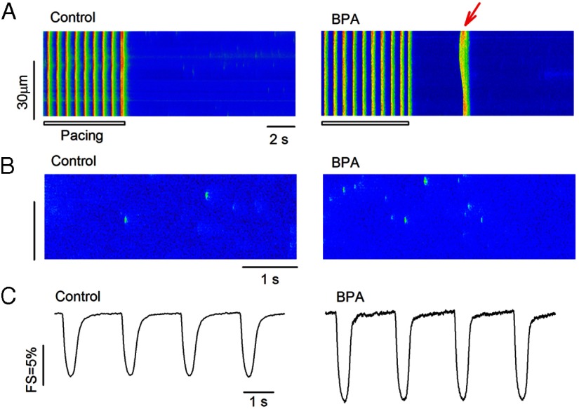
Rapid impact of BPA in female rat ventricular myocytes. A, Example confocal images showing Ca2+ transients in myocytes elicited by pacing under control (left panel) and upon acute exposure to 1 nM BPA (right panel). Arrow indicates spontaneous Ca2+ after-transient (ie, triggered activity) after pacing (n = 6 hearts). The control group had 49 myocytes, with three myocytes showing triggered activities. The BPA group had 52 myocytes, with 16 myocytes showing triggered activities (P < .01). Statistical analysis of the frequency of the triggered activities was performed using the χ2 test. B, Representative confocal images showing Ca2+ sparks in myocytes under control (left panel) and upon acute exposure to 1 nM BPA (right panel) (n = 3 hearts). Ca2+ spark frequency was 1.55 ± 0.18 (per 100 μm/sec) for control and 2.75 ± 0.19 (per 100 μm/sec) for the BPA treatment group (n = 12 myocytes for each group; P < .05). C, Representative contraction traces from myocytes under control (left panel) and upon acute exposure to 1 nM BPA (right panel; n = 4 hearts). FS was 8.3% ± 0.14% for control (n = 49) and 14.1% ± 0.21% for the BPA treatment group (n = 44 myocytes; P < .05). For B and C, statistical analysis was performed using a Student's t test. Data were expressed as average ± SEM.

