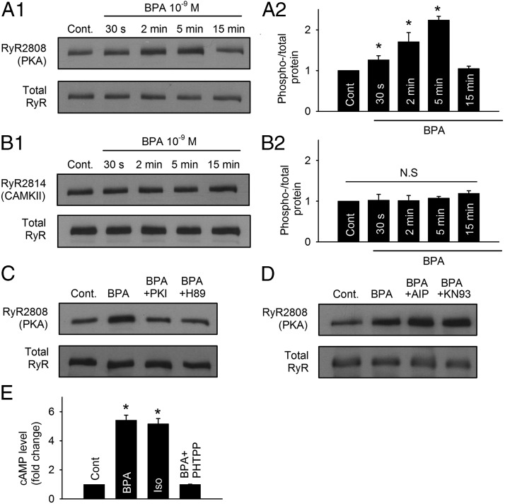
Figure 2.
Rapid impact of BPA on RyR via the cAMP/PKA pathway in female rat myocytes. A1 and A2, Representative immunoblot and quantification of RyR serine 2808 (PKA site) phosphorylation and total RyR under control and upon exposure to 1 nM BPA for indicated time points (n = 5 hearts). cont, control. B1 and B2, Representative immunoblot and quantification of RyR serine 2814 (CAMKII site) phosphorylation and total RyR under control and upon exposure to 1 nM BPA for indicated time points (n = 4 hearts). C, Immunoblot of RyR serine 2808 (PKA site) phosphorylation and total RyR expression under control, BPA, BPA + 1 μM PKI, and BPA + 1 μM H89 treatment for 5 minutes (n = 3 hearts). D, Immunoblot of RyR serine 2808 (PKA site) phosphorylation and total RyR expression under control, BPA, BPA + 1 μM AIP, and BPA + 1 μM KN93 treatment for 5 minutes. E, ELISA quantification of intracellular cAMP from myocytes under control and treated with BPA, 0.1 μM isoproterenol (Iso), and BPA + 5 μM PHTPP for 5 minutes (n = 3 hearts). All values were normalized to control. *, P < .05 vs control; N.S, not significant (P > .5).

