Figure 3.
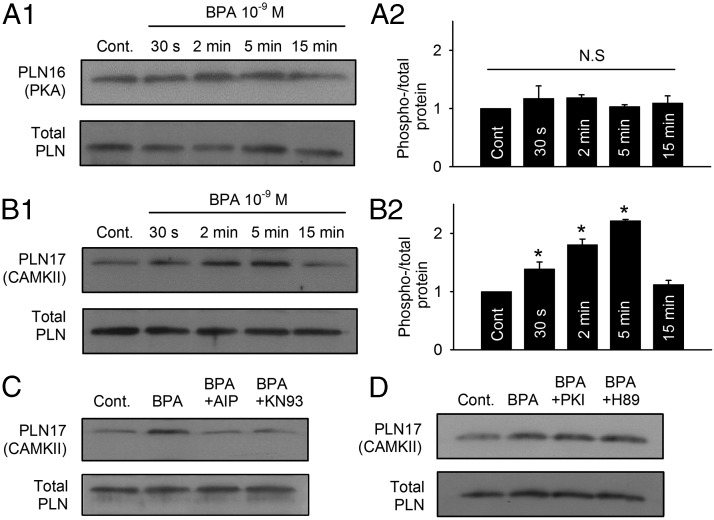
Rapid impact of BPA on PLN phosphorylation by CAMKII in female rat myocytes. A1 and A2, Representative immunoblot and quantification of PLN serine 16 (PKA site) phosphorylation and total PLN under control and upon exposure to 1 nM BPA for indicated time points (n = 3 hearts). cont, control. B1 and B2, Representative immunoblot and quantification of PLN threonine 17 (CAMKII site) phosphorylation and total PLN under control and upon exposure to 1 nM M BPA for indicated time points (n = 4 hearts). C, Immunoblot of PLN threonine 17 (CAMKII site) phosphorylation and total PLN under control, BPA, BPA + 1 μM AIP, and BPA + 1 μM KN93 treatment for 5 minutes (n = 4 hearts). D, Immunoblot of PLN threonine 17 (CAMKII site) phosphorylation and total PLN under control, BPA, BPA + 1 μM PKI, and BPA + 1 μM H89 treatment for 5 minutes. All values were normalized to control. *, P < .05 vs control; N.S, not significant (P > .5).

