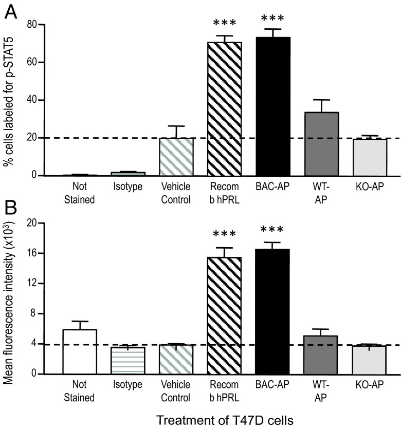
Figure 7.
Summary of flow cytometric analysis of STAT5 activation in human breast cancer T47D cells. See Supplemental materials for additional information. Flow cytometric analysis of fluorescent labeling measured the percent of T47D cells labeled (A) and the mean fluorescence intensity (degree of labeling) of those positive cells (B). The cells not stained or stained for κ isotype served as technical controls (2 bars on the left of each graph), whereas the remaining groups were all stained for phosphorylated STAT5. Vehicle-treated cells served as the biological control. Extracts of WT or mPRL−/− (PRL KO) APs did not increase either the percent of cells labeled or the mean fluorescence above vehicle treatment. Recombinant hPRL (10nM) increase both parameters approximately 4-fold over vehicle treatment and extracts of APs from hPRL+ mice (10nM hPRL, determined by RIA) were equipotent. Data represent the means of 4 repeated experiments. ***, P < .001 compared with vehicle control; 1-way ANOVA and Bonferroni's multiple comparison test.

