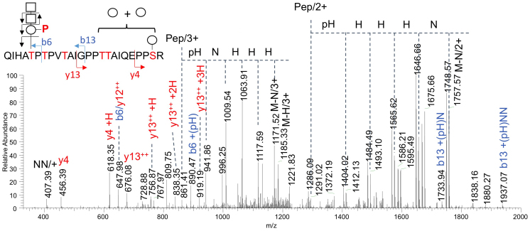
Figure 4. Determination of the presence of a phosphorylated trisaccharide on T317 of α-DG428 co-expressed with AGO61 in COS7 cells.
α-DG428 was purified using a HaloTag protein purification system from cell lysates co-transfected with α-DG428-HALO and AGO61, separated by SDS-PAGE, and in-gel digested with trypsin (Supplementary Fig. S6). The extracted peptides/glycopeptides were directly analyzed by LC-MS/MS. The successive neutral losses of HexNAc and PHex from both the doubly and triply charged molecular ions overlapped with those of the additional Hex and collectively defined the m/z of the bare peptide core with the fitted P1Hex4HexNAc2 glycosyl composition. The y4 + Hex and y13 + Hex1-3 fragment ions localized the additional 3 Hex on the C-terminal half of the peptide, whereas the b13 + PHex + HexNAc1-2 ions established the HexNAc2PHex substituent on the N-terminal half along with the b6 + PHex ion that further identified it on T317, as annotated. Key: Pep, peptide core; circle and H, Hex; square and N, HexNAc; P, phospho-; pH, phosphorylated Hex; M, molecular ion.

