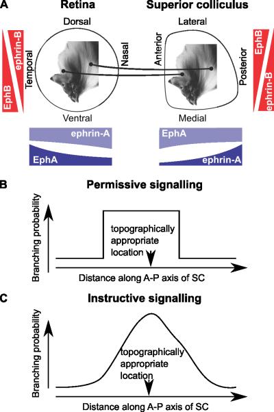
Figure 1. Retinocollicular mapping and permissive versus instructive molecular signalling.
A, Schematic diagram of retinocollicular mapping and forward and reverse gradients. Along the nasotemporal and anterior-posterior axes, the forward signalling system is made up of EphA in retina and ephrin-A in the superior colliculus (SC) and the reverse system comprises ephrin-A in the retina and EphA in the SC. The size of gradients is proportional to those used in the simulations shown in Figure 2. Along the retinal dorsoventral axis and the mediolateral axis of the SC EphB and ephrin-B make up the forward and reverse system; they are not modelled explicitly in Grimbert & Cang (2012), so the the gradients shown are purely schematic. B, Permissive cues restrict the region in which an axon can arborize, but do not favour any position within the permitted region. C, Instructive cues give the maximum arborization probability at the topographically appropriate location.

