
FIGURE 5.
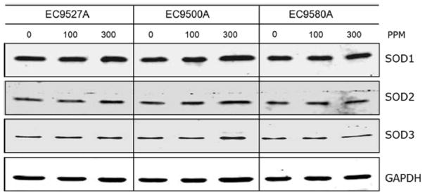
Superoxide dismutase levels following treatment with COREXIT dispersants. Western blot analysis was performed on cell extracts from BEAS-2B cells exposed to 0, 100, or 300 ppm dispersants for 2 h. Antibodies specific to superoxide dismutase proteins SOD1, SOD2, and SOD3 were used, with GAPDH was used as a loading control. Three independent experiments were conducted (a representative blot is shown).