
Figure 1.
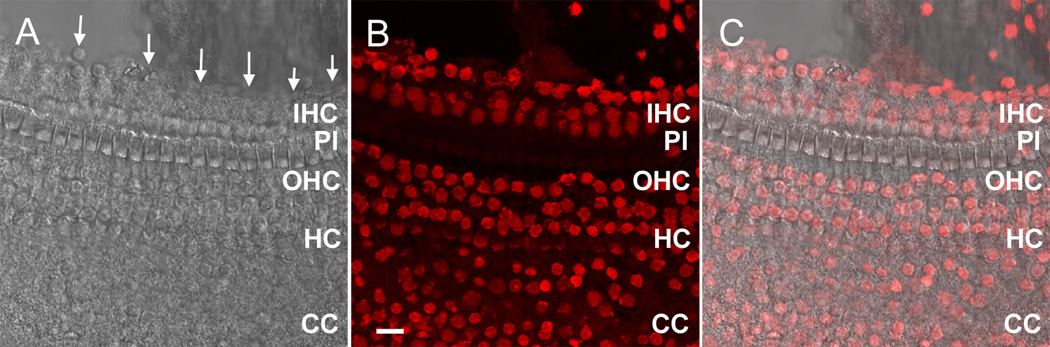
A microscopic view of the medial edge of the sensory organ partition following the initial cochlear preparation. A) A surface view of the tissue with arrows pointing to the medial edge. The osseous spiral lamina has been removed. B) The nuclei of the same tissue are stained with propidium iodide. The nuclei along the medial edge of the tissue (in the top section of the image) belong to IHCs, inner pillar cells, inner border cells and inner phalangeal cells. C) The merged image of (A) and (B). IHC: inner hair cell. PI: pillar cell. OHC: outer hair cell. HC: Hensen cell. CC: Claudius cell. Scale bar =15 µm.