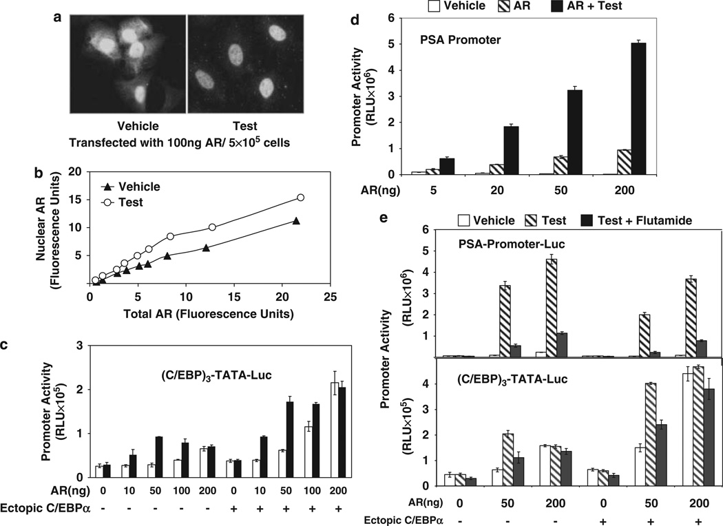
Figure 3.
The effect of forced nuclear localization of androgen receptor (AR) on ligand sensitivity of promoter activation by AR-CCAAT enhancer binding protein-a (C/EBPα). (a, b) Sub-cellular localization of ectopic AR in HeLa cells was examined using immunofluorescence. HeLa cells grown in chamber slides were transfected with different doses of AR expression plasmid (10–200 ng) or vector control and treated with either testosterone(10 nM) or vehicle for the duration of the transfection (48 h). Immunofluorescence staining for AR was performed using a primary rabbit antibody to AR and a bovine anti-rabbit immunoglobulin G (IgG)-fluorescein isothiocyanate (FITC) as the secondary antibody. The nuclei were stained with 4,6-diamidino-2-phenylindole (DAPI; not shown) and fluorescence images were captured using confocal microscopy. In (a), the representative images show a mixed distribution of AR (green fluorescence) between nuclear and cytosolic compartments in the absence of hormone but a predominantly nuclear distribution after testosterone treatment in the cells transfected with 100 ng AR expression plasmid per 5 × 10⁵ cells. In panel b, the total amount of AR fluorescence in the cell and the amount of AR fluorescence localized in the nucleus were quantified using LASAF software. The values are plotted as arbitrary fluorescence units. (c) HeLa cells were co-transfected with (C/EBP)3-TATA-Luc and different amounts of AR expression plasmid (10–200 ng per 5 × 10⁵ cells) or vector control together with C/EBPa expression plasmid or vector control. The cells were treated with testosterone (10 nM) or vehicle for the duration of the transfection (48 h) and harvested for luciferase assays. (d) HeLa cells were transfected with the prostate-specific antigen (PSA)-promoter luciferase reporter construct together with different amounts of AR expression plasmid (5–200 ng per 5 × 10⁵ cells). The cells were treated with testosterone (10 nM) or vehicle for the duration of the transfection (48 h) and harvested for luciferase assays. (e) HeLa cells were transfected with either PSA-promoter-Luc or (C/EBP)3-TATA-Luc and co-transfected with different amounts of AR expression plasmid (50–200 ng per 5 × 10⁵ cells) or vector control and C/EBPa expression plasmid or vector. The cells were treated with testosterone (10 nM), vehicle or the combination of testosterone (10 nM) and flutamide (25 µM) for the duration of the transfection (48 h) and then harvested for luciferase assays. For panels b-e, the P-values for the differences noted in the text were <0.001. A full colour version of this figure is available at the Oncogene journal online.

