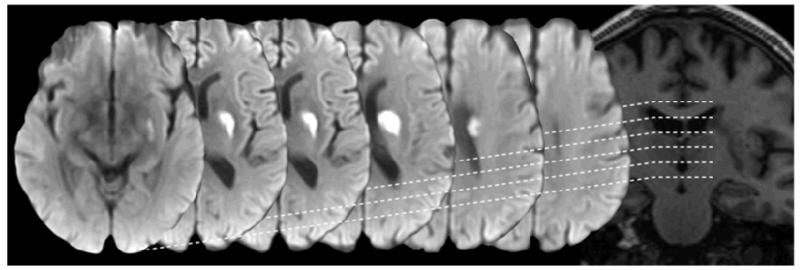
Figure 2.
An acute small deep (lacunar) infarct on diffusion imaging (serial axial views from basal ganglia to centrum semiovale, left to right) and T1-weighted imaging (coronal view, right). Note the tubular shape in the coronal plane as the infarct follows the line of a perforating arteriole. A wider range of examples of acute small deep (lacunar) infarcts is shown in Supplementary Figure 1.

