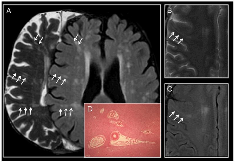
Figure 4.
Examples of perivascular spaces (PVS) on MRI and histology. (a) 72 year old asymptomatic subject, right, T2-weighted image shows linear PVS in the plane of the image, and on left FLAIR shows WMH around the PVS; (b) 49 year old man with left internal capsule acute small deep infarct (not shown) on T2-weighted imaging shows a perivascular space extending from the periventricular to subcortical tissues and (c) on the corresponding FLAIR image, one WMH running longitudinally around the PVS. (d) PVS on histology (H&E x40) showing parenchymal tissue retraction from around small perforating vessels; these have been dismissed as a processing artefact but are typically seen in ageing brain sections, and often associated with SVD.

