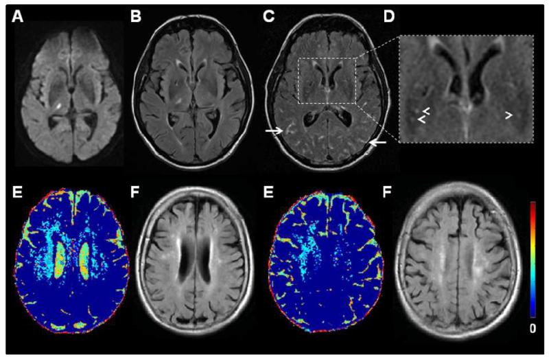
Figure 7.
MR imaging of cerebrovascular endothelial permeability. Top row: 56 year old patient with a right thalamic lacunar infarct: A) DWI, B) FLAIR two days after symptom onset. C) Two months later, FLAIR image after iv. gadolinium (Gd) showing Gd in the perivascular spaces (arrowheads) and sulci (arrows) and (D) inset magnified image of (C). Bottom row: Older patient with left internal capsule lacunar infarct (not shown): E) colour mapping of cerebrovascular permeability following intravenous Gd and F) corresponding FLAIR images showing WMH. Blue indicates low cerebral vascular endothelial permeability, yellow and red indicate increasing permeability. Permeability changes are diffuse. (E) courtesy of Dr Maria Valdes Hernandez.

