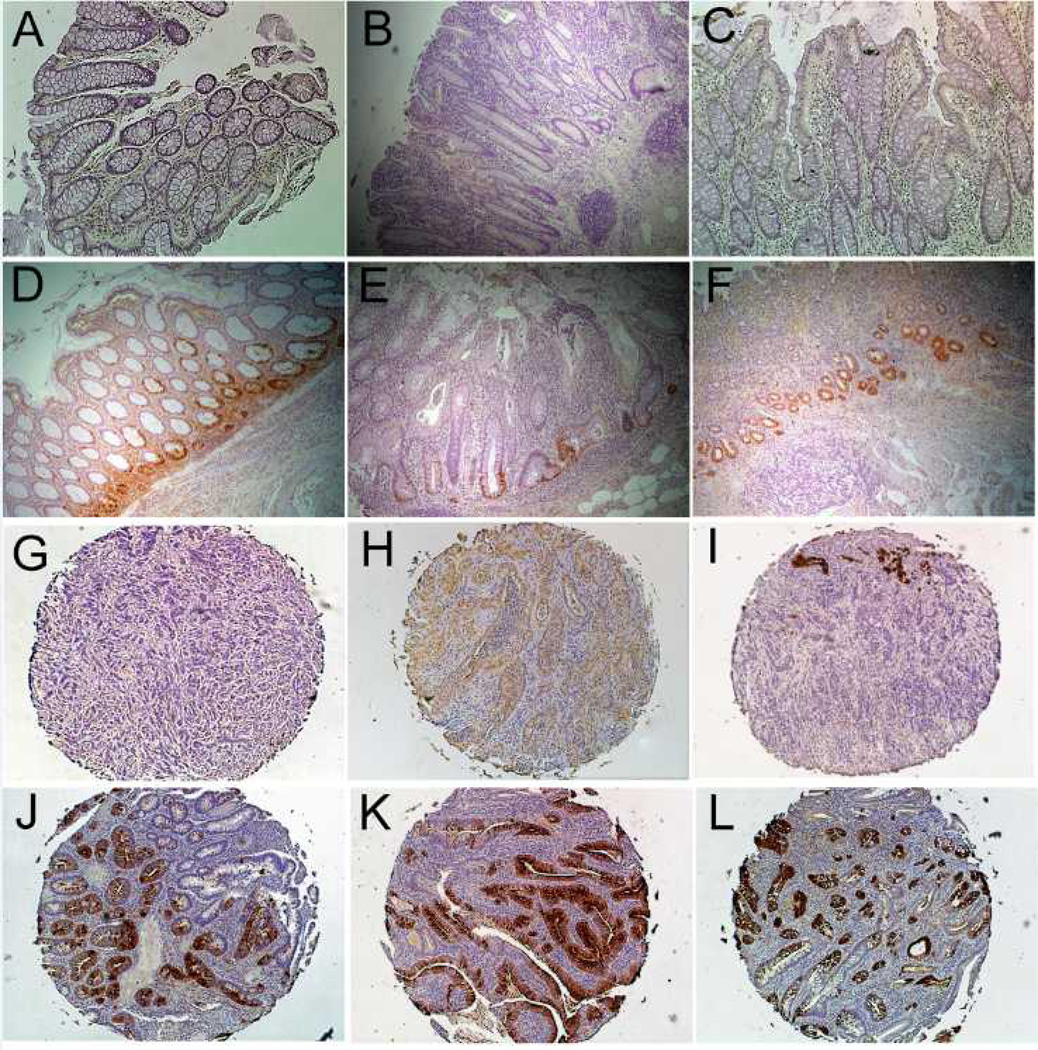
Figure 6.
REG1α Immunohistochemistry. (A) Normal colon, (B, C) UC without dysplasia, and (D–F) UC harboring a remote dysplastic lesion. In patients with UC harboring remote dysplasia, there is increased crypt staining, primarily in base of crypts. (G–L) IBD-cancer tissue array (G) A cancer with no positive staining, (H) Cancer with diffuse weak positive staining (I) A cancer with focal strong positive staining (J–L) Cancers with diffuse strong glandular positivity. Images A–G at 10x magnification. Images G–L at 5x magnification.

