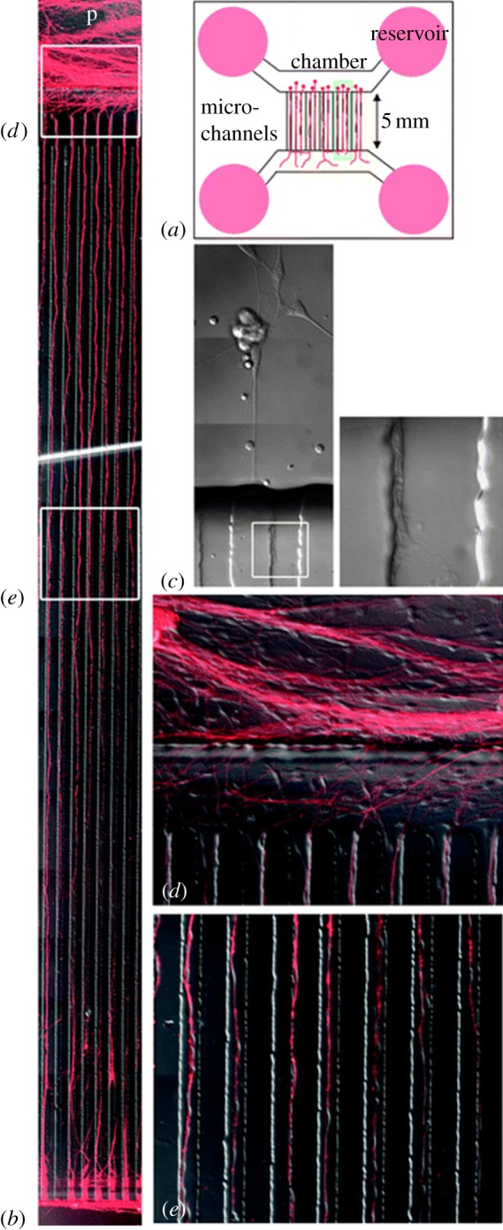
Figure 7.

(a) Schematic of the device can be seen, along with (b) representative images of DRG growth through microchannels, and a higher magnification of the same. (c) A representative axon bundle cut with a fem- tosecond laser demonstrates high thermal confinement of the site of injury. Axons can be seen (d) entering and (e) traveling through microchannels in higher resolution images (adapted from Kim et al. [88] with permission from the Royal Society of Chemistry).
