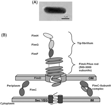
Figure 1.
Representations of type-1 fimbriae: (A) Electron micrograph of fimbriae on E. coli [38] and (B) the chaperone usher pathway.

Notes: Upon translation, subunits are secreted into the periplasm via the SecYEG translocon. FimC (the ‘chaperone’) then accelerates protein folding, and delivers the subunits to the pore forming protein FimD (‘the usher’) in the outer membrane. Here, the subunits are translocated and incorporated into the growing pilus.
