
Figure 5.
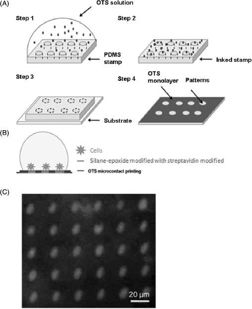
(A) Diagrammatic representation of the method used to pattern arrays of E. coli immobilised onto OTS functionalised surfaces (figures adapted from [96]). Step 1 involved the inking of the stamp with the OTS solution. The stamp was then dried with the application of a stream of nitrogen in step 2. Step 3 places the inked PDMS stamp onto the glass slide. The final step, step 4, shows that OTS molecules are transferred on the surface but not on the patterns that correspond to the carved structures of the PDMS stamp. (B) OTS μCP was followed by their modification with epoxide and streptavidin, followed by incubation with cells. (C) A fluorescence image of the selective binding of streptavidin molecules onto epoxide patterns, which allow the binding of the cells [96].