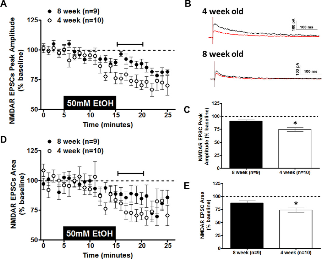
Figure 1. Effects of Acute Ethanol on NMDAR transmission in the BNST.
Acute ethanol (50 mm; 10 min) was applied to vBNST slices from 4- and 8-week-old C57BL/6J mice; evoked NMDA-receptor isolated EPSCs were then recorded. A) Time course of NMDAR-EPSC peak amplitude in 4- and 8-week-old mice. B) Representative traces of NMDAR-EPSCs before (black trace) and after (red trace) removal of ethanol in 4- and 8-week-old mice. C) Averaged peak amplitude of NMDAR-EPSC for the first 5 min after ethanol exposure in 4- and 8-week-old mice. D) Time course of NMDAREPSC area in 4- and 8-week-old mice. E) Averaged area of NMDAR-EPSC for the first 5 min after ethanol exposure in 4- and 8-week-old mice. *p < 0.05

