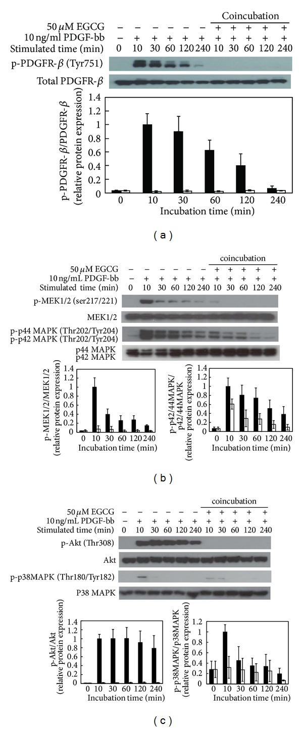
Figure 5.

The effect of EGCG on modulation of PDGF-bb stimulatory signal pathways in RAOSMC. Serum-starved RAOSMC was stimulated with 10 ng/mL PDGF-bb and 50 μM EGCG for the desired time (10 m, 30 m, 1 h, 2 h, and 4 h, resp.), lysed, and lysates were immunoblotted with antibodies. After densitometric quantification using the imageJ program, data were each expressed as the mean ± SD from three independent experiments. The black bar indicates expression by PDGF-bb stimulation. The white bar indicates expression by PDGF-bb stimulation with EGCG. (a) The expression of phospho-PDGFR-β (Tyr751) in a time-dependent manner. The band intensity was normalized to total PDGFR-β expression. (b) The expression of phospho-MEK1/2 (Ser217/221) and phospho-p42/44 MAPK (Thr202/Tyr204) in a time-dependent manner. The band intensity was normalized to total MEK1/2 and p42/44 MAPK expression. (c) The expression of phospho-Akt (Thr308) and phospho-p38 MAPK (Thr180/Tyr182) in a time-dependent manner. The band intensity was normalized to total AKt and p38 MAPK expression.
