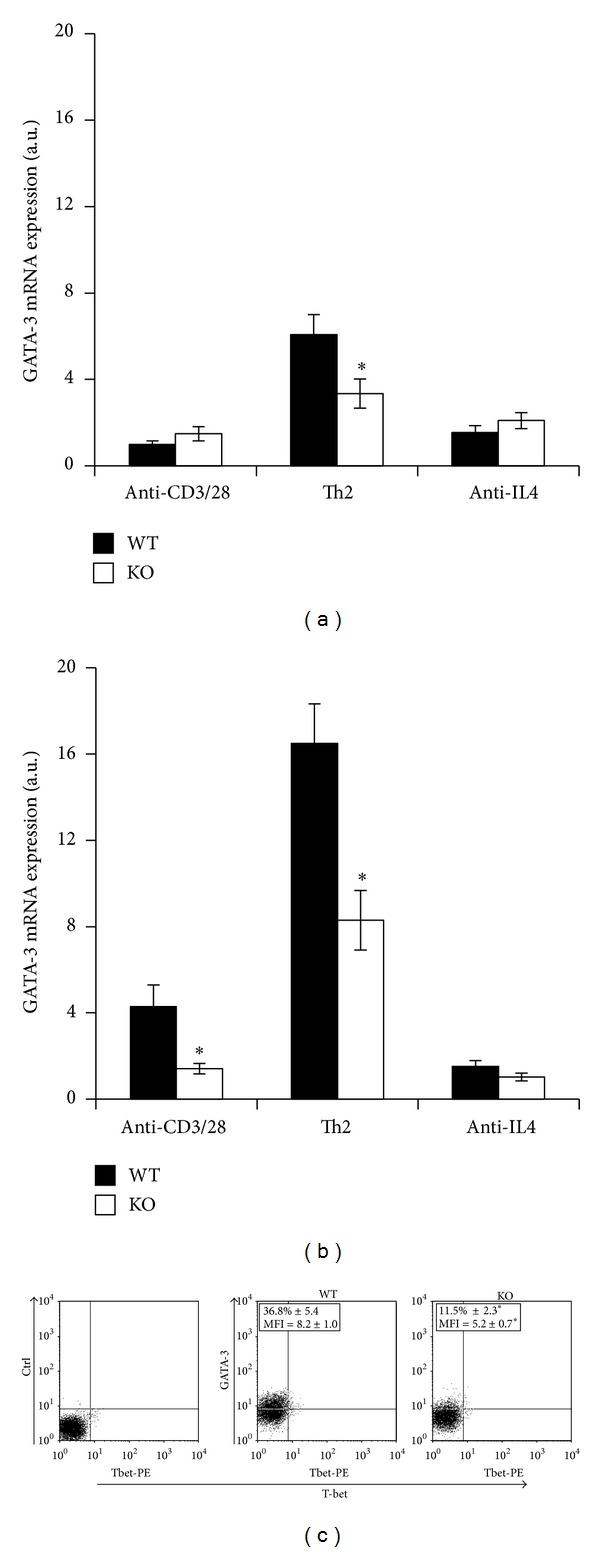
Figure 2.

PARP-1 deficient cells express GATA-3 at lower levels than WT cells. Naïve CD4 T cells from wild type (WT, filled symbols) and PARP-1KO (KO, unfilled symbols) mice were stimulated with anti-CD3 and anti-CD28 mAbs alone or together with either IL-4 and neutralizing anti-IFN-γ mAb (Th2-polarzing conditions) or anti-IL4 neutralizing mAb (anti-IL4). GATA-3 mRNA expression was assessed by reverse transcription and real time PCR after 24 (a) and 72 (b) hours of stimulation. Results, normalized on the housekeeping gene (GAPDH) mRNA level, are referred to the GATA-3 mRNA level in WT cells stimulated for 24 hrs and expressed as arbitrary units. Values represent means ± S.E. from three independent experiments. (c) After 72 hours of stimulation with anti-CD3, rCD86 cells and IL-4 (and anti-IFN-γ) cells were analyzed by flow cytometry. Representative dot plots are shown. The indicated percentage of GATA-3-expressing cells and mean fluorescence intensity (MFI) for GATA-3 staining are means (±S.E.) from three independent experiments. *P < 0.05 for KO versus WT cells, where indicated in (a), (b), and (c).
