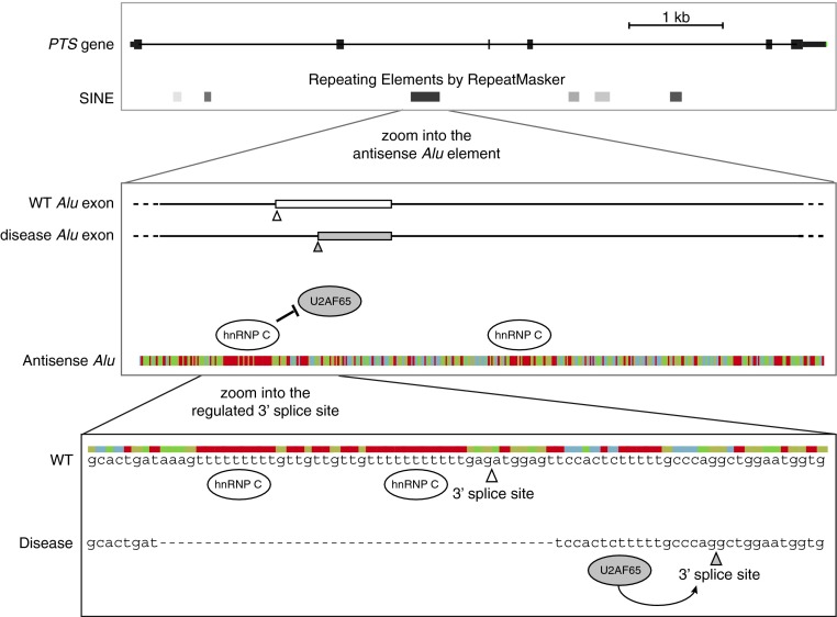
Figure 1. hnRNP C repression of Alu exonization in the PTS gene is relevant for disease.
(A) A view of the exon/intron structure of the PTS gene. The boxes below show the positions of short interspersed elements (SINE), which include Alu elements, as defined by RepeatMasker. (B) The disease-relevant Alu element within the PTS gene is shown at greater resolution. The position of the two exons that can emerge from this Alu element are schematically indicated: the blank exon is rarely included in the wild-type (WT) cells, whereas the grey exon is part of the dominant isoform in disease. Below, the sequence is shown in a colour-coded fashion. The uridine tracts (in red) bind to hnRNP C, which represses binding of U2AF65. (C) The two 3′ splice sites that can lead to the formation of Alu exon are shown at nucleotide resolution. In wild-type cells, hnRNP C binds to the long uridine tracts to repress the binding of U2AF65, and therefore the 3′ splice site marked by the open arrowhead is rarely used. In disease, the primary hnRNP C-binding site is deleted, and therefore U2AF65 can bind to the pyrimidine tract upstream of the 3′ splice site that is marked by the grey arrowhead. This leads to strong inclusion of the grey exon that is shown in (B) [8].

