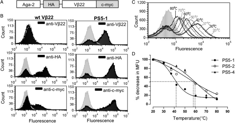
Fig. 1.
Yeast surface display of human Vβ22. (A) Schematic of yeast display construct containing human Vβ22 gene. (B) Flow cytometric analysis of induced yeast cells expressing either wt Vβ22 or PS5-1 mutant protein. Yeast cells were stained with anti-Vβ22, anti-HA or anti-c-myc (black) followed by a secondary antibody conjugated to a fluorophore. The staining profile of the cells with secondary antibody only is represented by the gray peak. Data shown are representative of three or more experiments. (C) Flow cytometric analysis of the stabilized Vβ22 mutant PS5-1 after yeast cells were incubated at various temperatures for 30 min prior to incubation with primary antibody (anti-Vβ22). (D) Thermostability analysis of stabilized Vβ22 mutants (PS5-1, PS5-2 and PS5-4) by flow cytometry, as described in (C). The point of the 50% melting temperatures of individual mutants are indicated.

