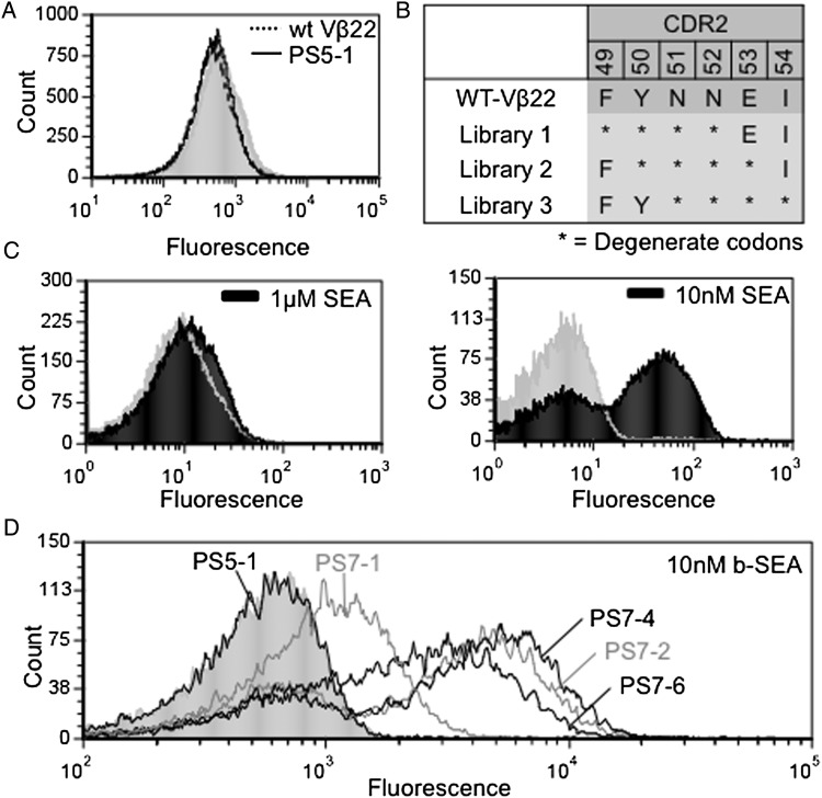
Fig. 3.
Generation of Vβ22 mutants that bind SEA with higher affinity. (A) Flow cytometry histograms for wt Vβ22 (dotted line) and PS5-1 (solid line) with 2.5 μM biotinylated-SEA followed by Streptavidin-Alexa 488. (B) Residue positions of three site-directed mutagenesis CDR2 libraries used to select higher affinity SEA binders. (C) Flow cytometry histograms of a population of Vβ22 mutants after sorting the combined CDR2 libraries with 10 nM biotinylated-SEA (right), compared with unsorted CDR2 libraries stained with 1 μM biotinylated-SEA (left). (D) Flow cytometry histograms for individual clones (PS7-1, PS7-2, PS7-4 and PS7-6) isolated after the seventh sort of the Vβ22 CDR2 libraries, incubated with10 nM biotinylated-SEA. Fluorescence with secondary reagent only (the absence of SEA) are represented by gray (filled) peak in each histogram. Data shown are representative of three experiments.

