Fig. 1.
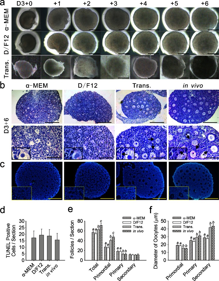
Morphology of incubated ovaries and the development of follicles. a Observations of in-vitro-cultured and transplanted (Trans.) ovaries by stereomicroscopy (D3+0 time at which the ovaries were dissected from the 3-day-old mice). Images were captured every 24 h. Ovaries appeared healthy, without any obvious dark areas. b Ovaries were cultured for 6 days in enriched α-MEM or enriched D/F12, transplanted for 6 days under the kidney capsule in castrated mice and then freshly isolated from 9-day-old mice. Hematoxylin/eosin (H&E) staining (arrows thecal layer). Bars 160 μm (top row), 80 μm (bottom row). Insets High-magnification views. c TUNEL assays with no observable differences in cellular apoptosis among the four groups. Bars 80 μm. Insets High-magnification views. d Numbers of positively stained cells for TUNEL were counted in each stained section. Values are expressed as the means ± SEM; n = 11–15. e Following culture and transplantation, follicles were classified and quantified. Values are expressed as the means ± SEM; n = 20–22. Values with different letters were significantly different in the same stage follicles from the four groups (P < 0.05). f Mean diameters (μm ± SEM) of the oocytes from follicles at various stages in the cultured, transplanted (Trans.) and in-vivo-grown ovaries. Values with different letters were significantly different in the same stage follicles taken from the four groups (P < 0.05); n = 20–22

