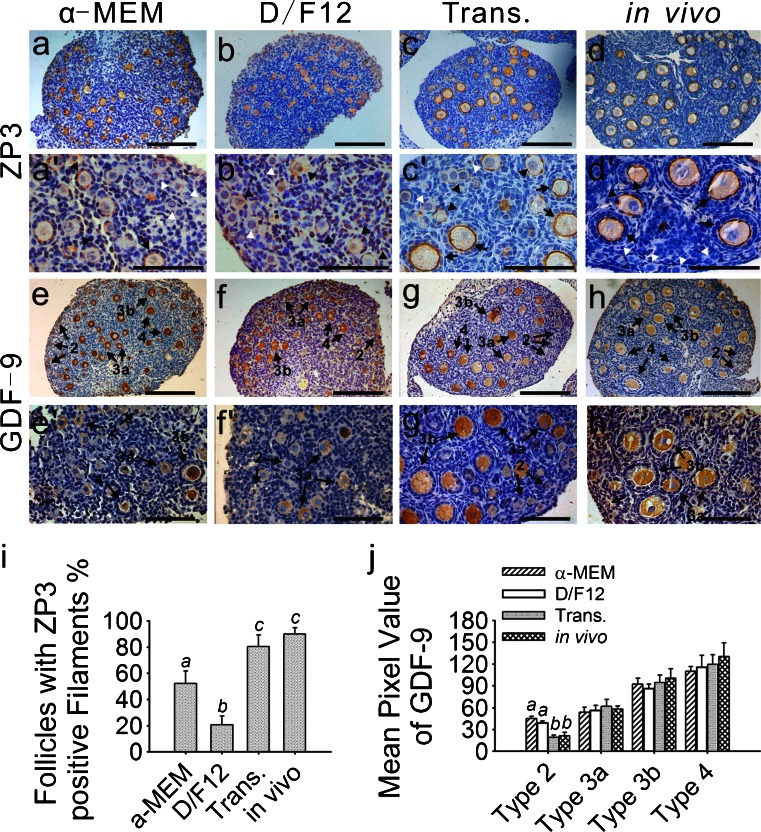
Fig. 2.
Immunohistochemical localization of zona pellucida 3 (ZP3) and growth and differentiation factor-9 (GDF-9). Ovaries that were freshly isolated from 9-day-old mice, cultured or transplanted for 6 days were collected. Immunohistochemistry was performed as described. a-d, a’-d’ ZP3 expression was present in all of the developing follicles in the four groups, whereas no staining was observed in the primordial follicles (white arrowhead). Compared with the transplanted (c, c’) and in-vivo-grown ovaries (d, d’), the developing follicles from the ovaries cultured in enriched α-MEM (a, a’) contained intact but thinner ZP3-positive filaments (arrows). Staining in the D/F12-medium-cultured ovaries was predominantly located in the cytoplasm of the oocytes (arrowheads follicles lacking intact filaments) and lacked an intact filamentous structure surrounding the oocytes (b, b’). Strongly ZP3-positive filaments were observed in most of the developing follicles (arrows) from the transplanted (c, c’) and in-vivo-grown ovaries (d, d’). e, f, e’, f’ GDF-9 staining was specific to the cytoplasm of the oocytes. In the cultured ovaries (e, f, e’, f’), the oocytes from the type 2 follicles began to demonstrate weak staining in the cytoplasm and more intense staining was detected in the more developed follicles. However, no immunoreactivity was present in the type 2 follicles in the transplanted (g, g’) or in-vivo-grown ovaries (h, h’). Bars 160 μm (a–h), 80 μm (a’–h’). i Percentages of follicles with intact zona pellucida filaments are represented as the percentage of total developing follicles from each group. In the ovaries cultured in enriched D/F12, only 20.7 % of the developing follicles contained ZP3-positive filamentous structures; this was significantly lower than the value in the other groups. For example, the ovaries cultured in enriched α-MEM contained 52.1 % developing follicles with ZP3-positive filamentous structures. No significant difference was observed between the transplanted (80.3 %) and in-vivo-grown ovaries (90.0 %). Different letters indicate significant differences among the four groups (P < 0.05); n = 19. j Intensity of the GDF-9 immunoreaction was measured as the mean value of pixel brightness in the cytoplasm of the oocytes. Follicles were classified as described. Higher pixel value reflects higher immunoreactivity. In cultured ovaries, the mean pixel value of the type 2 (primordial) follicles was significantly high. No significant difference was seen in the other stages of follicles from the four groups. Different letters indicate significant differences among the follicles at the same stage taken from the four different groups (P < 0.05); n = 20

