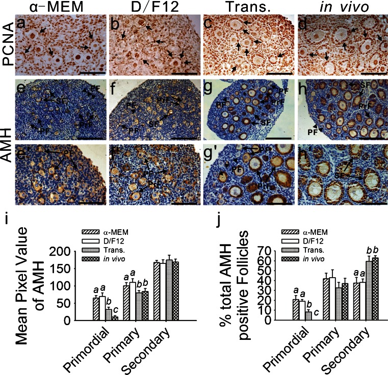
Fig. 3.
Immunohistochemical localization of proliferating cell nuclear antigen (PCNA) and anti-Müllerian hormone (AMH). a–d PCNA expression was specific to the nuclei of both the oocytes and granulosa cells. To reveal clear PCNA-positive staining, the nuclei were not counterstained with hematoxylin. Positive immunoreactivity (arrows) is represented by dark brown staining. e-h, e’–h’ AMH immunoreactivity was present in the cytoplasm of the granulosa cells. The theca cells and interstitium in all of the ovaries failed to stain for AMH. In the cultured (e, f, e’, f’) and transplanted ovaries (g, g’), AMH expression was found in the granulosa cells from some of the primordial, primary and secondary follicles (PF primordial follicle, PrF primary follicle, SF secondary follicles, arrow positive follicles, arrowhead negative follicles). No AMH-positive primordial follicles were found in the in-vivo-grown ovaries (h, h’). Bars 160 μm (a–h), 80 μm (e’–h’).i Intensity of the AMH immunoreaction was measured as the mean value of pixel brightness in the granulosa cells of the follicles at various stages. Higher pixel value represents higher immunoreactivity. Different letters indicate significant differences among the mean values of pixel brightness in the granulosa cells of the same stage follicles from the four different groups (p < 0.05); n = 21. j Percentages of AMH-positive follicles at different stages are represented as the % of total AMH-positive follicles from each group. Different letters indicate significant differences among the percentages of positive follicles at the same stage taken from the four different groups (P < 0.05); n = 21

