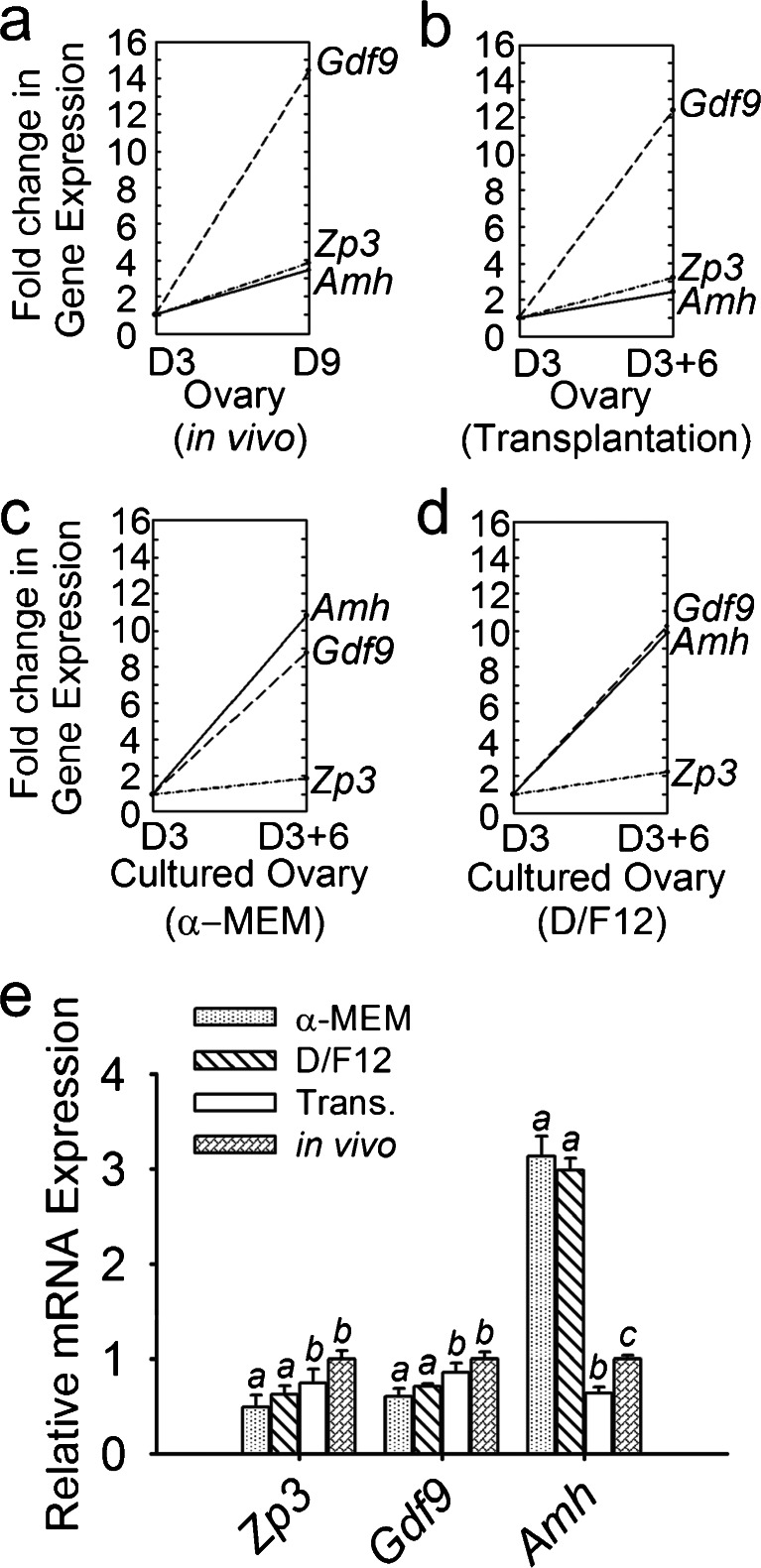
Fig. 4.
Relative gene expression patterns and levels in the four groups. Gene expression levels of the ovaries that were freshly isolated from 3-day-old mice were defined as the baselines. Compared with each baseline, the fold changes are presented for the freshly isolated ovaries from 9-day-old mice (a), ovaries incubated under the kidney capsule of castrated mice for 6 days (b), ovaries cultured for 6 days in enriched α-MEM (c) and ovaries cultured for 6 days in enriched D/F12 (d). The fold changes in the expression of Zp3, Gdf9 and Amh among these four groups are shown in e. In the in-vivo-grown ovaries, the expression of Zp3, Gdf9 and Amh in freshly isolated 9-day-old ovaries increased, with fold changes of 3.43, 14.47 and 3.70, respectively, compared with 3-day-old ovaries. Amh significantly increased approximately 11-fold after 6 days in culture and decreased after transplantation (Trans.; P < 0.05). Compared with the in-vivo-grown ovaries, the fold changes in the expression of Zp3 and Gdf9 were lower in the cultured ovaries (P < 0.05), whereas they were similar in the transplanted ovaries (P > 0.05). Data are presented as the means ± SEM from five separate experiments; n = 7 per group. Values with different letters denote significant differences (P < 0.05)

