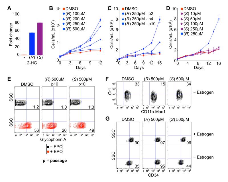
Fig. 2.
(R)-2-HG is sufficient to promote leukemogenesis. (A) LC-MS analysis of TF-1 cells after treatment for 3 hours with DMSO (-) or 250 μM of the indicated esterified (TFMB) 2-HG. Shown are mean values of triplicate experiments. (B-D) Proliferation of TF-1 cells under cytokine-poor conditions in the presence of the indicated amounts of TFMB-(R)-2-HG (B and C) or TFMB-(S)-2-HG (D). Cells in (B and D) were passaged 10 times prior to GM-CSF withdrawal. Cells treated with 250 μM TFMB-(R)-2-HG are also included in (D) for comparison. Shown are mean values of duplicate experiments ± SD. (E) Differentiation of TF-1 cells, as determined by Glycophorin A FACS, after 8 days of EPO treatment following pretreatment for 10 passages with DMSO or 500 μM TFMB-2-HG (R or S). Shown are representative results of three independent experiments. (F and G) Differentiation of SCF ER-Hoxb8 cells, as determined by dual staining for CD11b/Mac1 and Gr1 (F) or staining for CD34 (G), 3 days after of withdrawal of β-estradiol following pretreatment with DMSO or 500 μM TFMB-2-HG (R or S) for 20 passages. Shown are representative results of two independent experiments.

