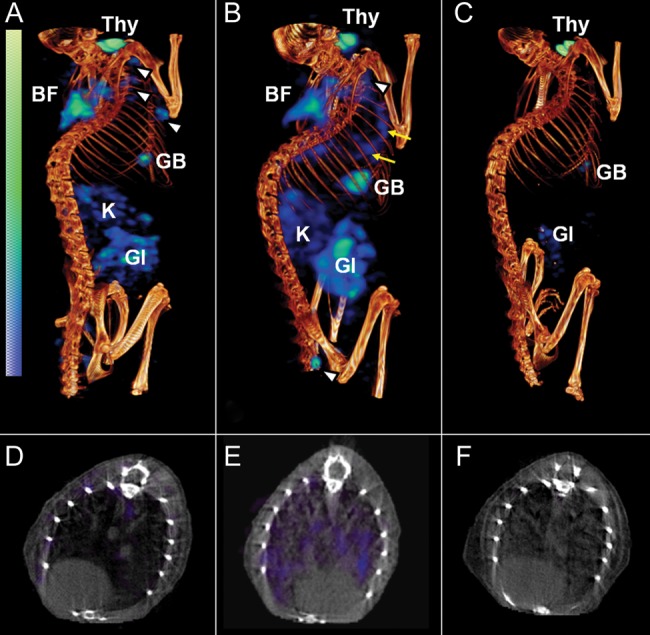
Figure 1.

[125I]Iodo-DPA-713 single-photon emission computed tomography–high-resolution CT (SPECT-CT) of BALB/c mice. Coregistered [125I]iodo-DPA-713 SPECT-CT images from representative Mycobacterium tuberculosis–infected BALB/c mice are shown. Compared with uninfected control mice (A and D), in which only a minimal pulmonary SPECT signal is noted, significant but diffuse signal is observed in the lungs of infected mice (B and E; yellow arrows). Auto-blockade abrogates the SPECT signal from the lungs of infected mice (C and F). Signal is also noted in brown fat (BF), lymph nodes (white arrowheads), gastrointestinal tract (GI), kidneys/adrenal glands (K), gallbladder (GB), and thyroid (Thy).
