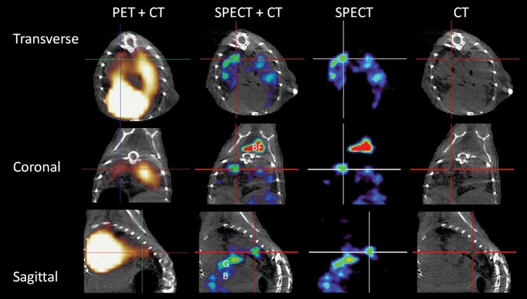
Figure 3.
[125I]Iodo-DPA-713 localizes to tuberculosis lesions. The transverse, coronal, and sagittal images from a representative Mycobacterium tuberculosis–infected C3HeB/FeJ mouse that underwent both [125I]iodo-DPA-713 single-photon emission computed tomography–high-resolution CT (SPECT-CT) and [18F]FDG positron emission tomography (PET)–high-resolution CT are shown. Discrete areas of [125I]iodo-DPA-713 SPECT signal are noted in the lungs of the infected mouse. The SPECT signal colocalizes with the tuberculosis lesion seen on CT (crosshair). The [18F]FDG PET signal is more diffuse, with intense signal noted in the heart, obscuring the pericardiac regions. [125I]iodo-DPA-713 SPECT signal is also noted in the brown fat (BF) in the coronal view.

