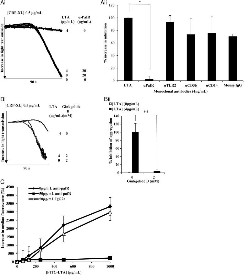
Figure 4.
LTA acts through PafR to inhibit platelets. A, Washed platelets were incubated with anti-PafR (4 µg/mL), anti-TLR2 (4 µg/mL), anti-CD14 (4 µg/mL), Mouse IgG (4 µg/mL) or Tyrodes buffer for 30 minutes before addition of LTA (4 µg/mL) for 15 minutes. B, Washed platelets were incubated with Ginkgolide B (2 mM) or Tyrodes buffer for 30 minutes before the addition of LTA (4 µg/mL) for 15 minutes. Platelets were then stimulated with CRP-XL (0.5 µg/mL). NB: In Bi, lines representing platelets treated with 4 µg/mL LTA + 2 µM Ginkgolide B and 0 µg mL−1 LTA + 2 µM Ginkgolide B, overlap extensively. A and B, Platelet aggregation was measured as change in light transmission and recorded for 90 seconds. Data are plotted as percentage inhibition of aggregation (normalized so that LTA treatment represents 100% inhibition) and represent mean values ± SEM. *P < .0001, **P < .01. C, PRP (4 × 10⁸ cells/mL) was incubated for 30 minutes with anti-PafR (50 µg/mL), IgG2a (50 µg/mL) or Tyrode buffer before the addition of FITC-LTA at several concentrations for 15 minutes. Samples were run through a BD Accuri C6 flow cytometer and median fluorescence was recorded. Data are plotted as percentage median increase in fluorescence when compared to a Tyrode buffer only control and represent mean values ± SEM. Abbreviations: CRP-XL, cross-linked collagen-related peptide; IgG, immunoglobulin G; LTA, lipoteichoic acid; PafR, platelet activating factor receptor; SEM, standard error of the mean.

