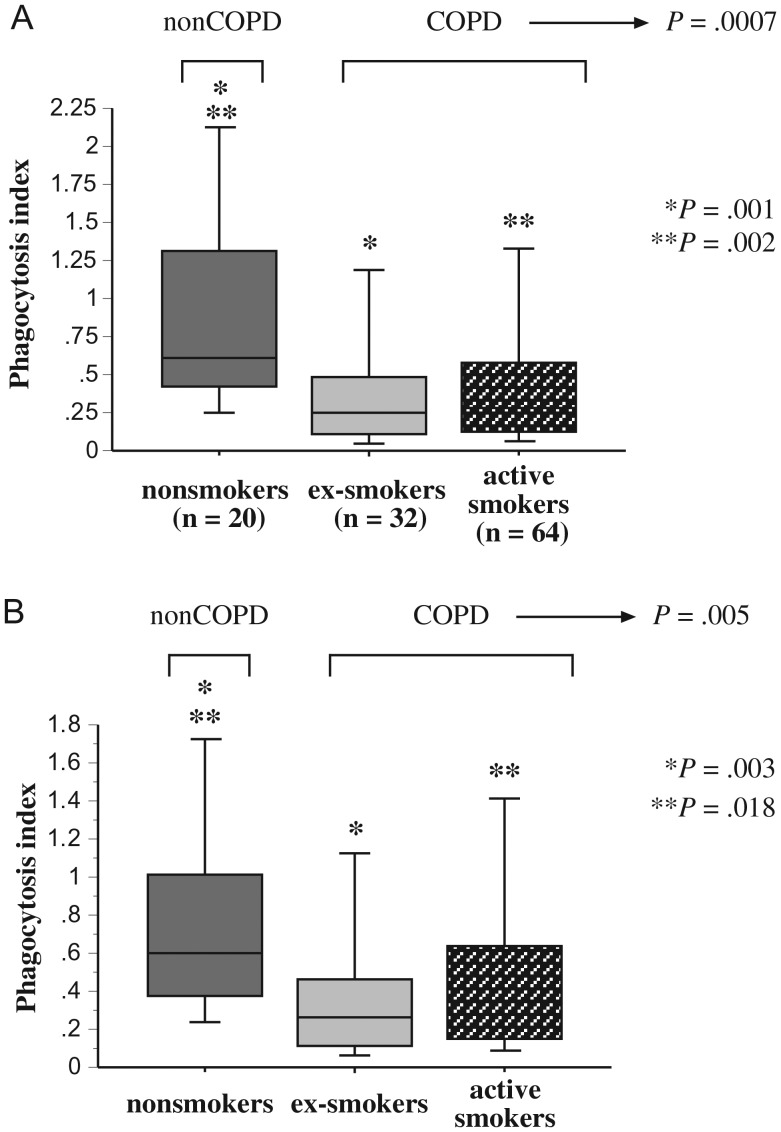
Figure 2.
Phagocytosis of Moraxella catarrhalis by human alveolar macrophages. Alveolar macrophages were obtained from nonsmokers, ex-smokers with COPD, and active smokers with COPD. Shading denoting each individual group is as described in Figure 1. Cells were incubated with M. catarrhalis 6P29B1 with complement (A) or in serum-free media (B). Phagocytosis was diminished for alveolar macrophages of COPD ex-smokers vs healthy nonsmokers (A: *P = .001; B: *P = .003) and for COPD active smokers vs healthy nonsmokers (A: **P = .002; B: **P = .018). Phagocytosis among all COPD participants was significantly less than that of healthy nonsmokers (A: P = .0007; B: P = .005). Data are represented by box plots for each group, as detailed in Figure 1. Data correspond with values of Supplementary Table 2. Statistical comparison of all 3 groups was performed by Kruskal-Wallis test and for intergroup comparisons by Mann–Whitney U rank test.

