
Fig. 4.
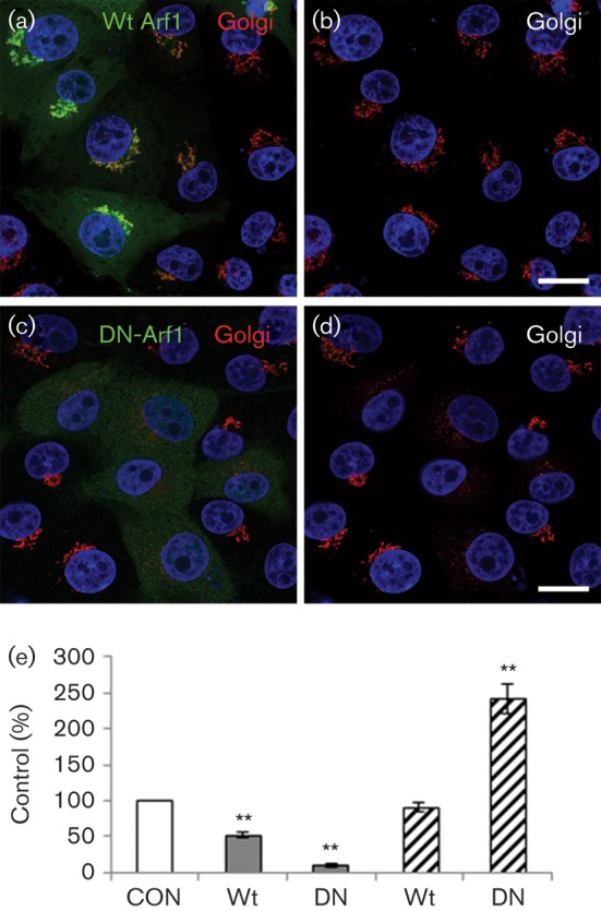
DN-Arf1 enhances infection by FMDV but not BEV. IBRS2 cells (on coverslips) were transfected with either wt Arf1 (GFP-wt-Arf1) or DN-Arf1 (HA-DN-Arf1T31N). (a) Cells transfected with GFP-wt-Arf1 (green) labelled for the Golgi (GM130, red). (b) The same cells as in (a) showing Golgi labelling only. (c) Cells transfected with HA-DN-Arf1T31N labelled for the HA tag (green) and Golgi (GM130, red). (d) The same cells as in (c) showing Golgi labelling only. The cell nuclei are shown in blue. Bars, 10 μm. (e) Cells were transfected with GFP-wt-Arf1 (wt) or HA-DN-Arf1T31N (DN) and infected with BEV (grey bars) or FMDV (hatched bars) (m.o.i. 0.3) for 3.5 h. The level of infection of cells positive for an Arf1 transgene was normalized to the non-expressing cells (control, CON) of the same coverslip. The data show the mean±sem for three independent experiments, each carried out with triplicate samples (n≥500 cells per coverslip). Student’s t-test was used to determine statistical significance (**P<0.01).