
Fig. 5.
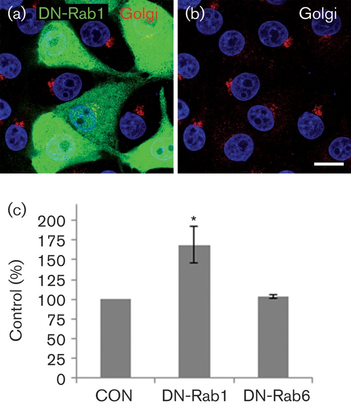
FMDV infection is enhanced by DN-Rab1. IBRS2 cells were transfected with either DN-Rab1a (myc-DN-Rab1aS25N) or DN-Rab6 (myc-DN-Rab6T27N). (a) Cells transfected with myc-DN-Rab1aS25N labelled for the myc tag (green) and the Golgi (giantin, red). (b) The same cells as in (a) showing Golgi labelling only. The cell nuclei are shown in blue. Bars, 10 μm. (c) Cells transfected with myc-DN-Rab1aS25N or myc-DN-Rab6T27N were infected with FMDV (m.o.i. 0.5) for 3.5 h. The level of infection of cells positive for a DN-Rab transgene was normalized to the non-expressing cells (control, CON) of the same coverslip. The data show the mean±sem for two independent experiments, each carried out with triplicate samples (n≥500 cells per coverslip). Student’s t-test was used to determine statistical significance (*P<0.05).