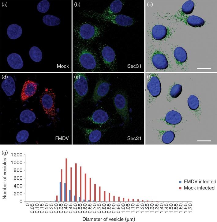
Fig. 8.
FMDV infection leads to dispersal and reduction of Sec31 labelling. HeLa cells were mock infected or infected with FMDV O1BFS/1860 (m.o.i 0.5) for 3 h. (a, b) Cell nuclei (a) and Sec31 labelling (green) (b) in mock-infected cells. (c) A rendered image of the cells shown in (b). (d) FMDV (labelled for 3A; red) and (e) Sec31 labelling (green) in infected cells. (f) A rendered image of the cells shown in (e). The cell nuclei are shown in blue. Bars, 10 μm. The IMARIS spot function was used to identify Sec31-positive punctae in 35 mock-infected and 35 infected cells. (g) Sec31 punctae size plotted against frequency.

