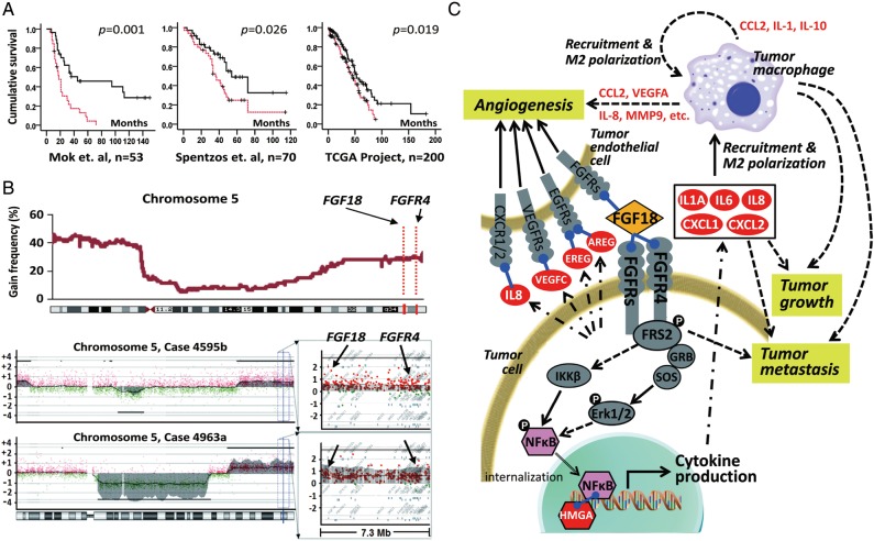
Figure 4.
Identification of FGF18 as a prognostic gene in high-grade advanced-stage papillary serous ovarian tumors. (A) Kaplan–Meier analysis of FGF18 expression in patients in three independent sets of serous ovarian cancer samples. Analysis was done by median cut with the P-value of log-rank test presented for each set. Solid lines: samples with low FGF18, broken lines: samples with high FGF18, ‘+’: censored samples. (B) CGH analysis of 72 serous ovarian tumors showed an increased DNA copy number for chromosome segment 5q31.3–5q35.3 in ∼25% of the samples (upper panel). Chromosome 5 profiles of two representative tumors with the detail of a 7.3-Mb locus (chromosome 5 distance: 170.3–177.6 Mb) containing FGF18 and FGFR4 (lower panel). Copy number was presented by log2 − 1 (value of 0 mean diploid). FGF18 and FGFR4 are amplified to at least four copies among the 5615 probes in the x-axis. (C) The effect of FGF18 on the pathogenesis of serous ovarian cancer. Gene expression profiling was carried out to compare the transcriptome of ovarian cancer cell lines with ectopic overexpression of FGF18 or RFP as control. Genes directly induced by FGF18 are labeled in white font. Arrows indicate potential interactions that contribute to FGF18-mediated ovarian tumorigenesis. Adapted from Wei et al.

