Figure 4.

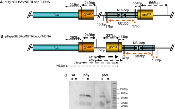
A and B. HpaII/MspI restriction map of gpdII promoters in pHg/pSILBAγ/NITRLoop (A) and pHg/pSILBAα/NITRLoop (B) T‐DNAs. CCGG restriction sites of HpaII (CpG methylation sensitive) and MspI (CpG methylation insensitive) within and around the promoter sequences are marked with vertical lines. Fragments from complete DNA digestion hybridizing to gpdIIP probe are indicated by arrows above the T‐DNAs. Detected fragments are marked with thicker arrows. Fragments from incomplete digestion of pHg/pSILBAα/NITRLoop with HpaII are indicated by black arrows below the T‐DNA in (B). Red arrows correspond to fragments from complete digestion of the nitrate reductase silencing trigger loop (not revealed in the Southern). The terminator elements of the T‐DNAs are not presented. C. Southern blot hybridized to gpdIIP probe. Results of one strongly silenced pSα, one strongly silenced pSγ and wild‐type strains are shown. If no CpG methylation is present in the target DNA both HpaII and MspI restrictions should result in identical hybridization signals. H: HpaII restriction. M: MspI restriction.
