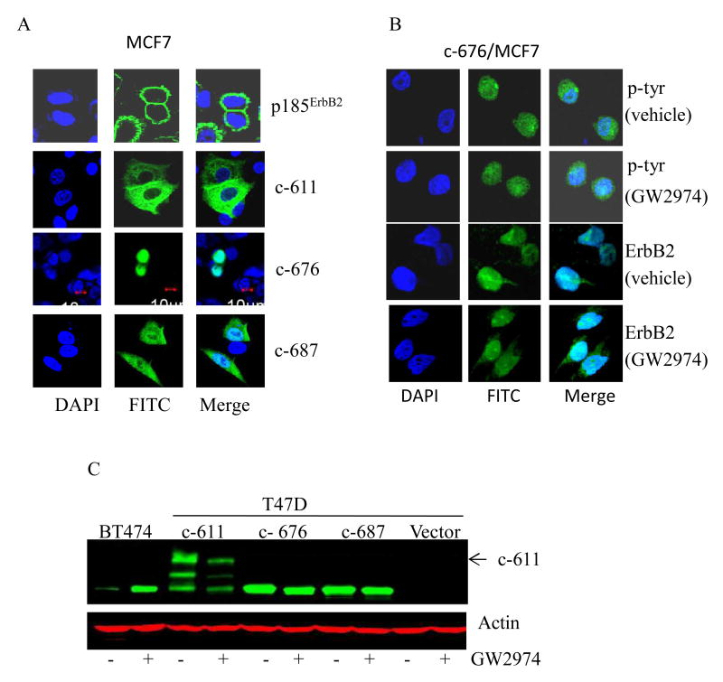
Figure 3. Similarity of p95L induced by ErbB2 TKI and ErbB2 c-terminal fragments (CTFs) generated by alternate translation initiation.
(A) The subcellular localization of the indicated CTF (c-611; c-676; c687) and p185ErbB2 expressed in MCF7 cells was determined by IF microscopy using an ErbB2 specific antibody and a FITC-conjugated secondary antibody (green). Nuclei were counterstained blue with DAPI and the overlap of DAPI and FITC is shown (Merge). (B) MCF7 cells expressing c-676 were treated for 24 h with GW2974 (8 μM) or vehicle alone (DMSO). The subcellular localization of p-tyr and c-676 signals was determined by IF microscopy using phosphotyrosine (p-tyr) and ErbB2 specific antibodies, respectively, and visualized with a FITC-conjugated secondary antibody (green). The overlap between DAPI and FITC is shown (Merge). (C) Phospho-ErbB2 steady-state protein levels (green) were determined by Western blot in T47D cells transfected with the indicated CTF's following treatment with GW2974 (8 μM) or vehicle (DMSO) alone for 24 h. Actin (red) steady-state protein levels served as a control for equal loading of protein. The arrow indicates the mature form of c-611. Results shown in Figure 3 are representative of three independent experiments.

