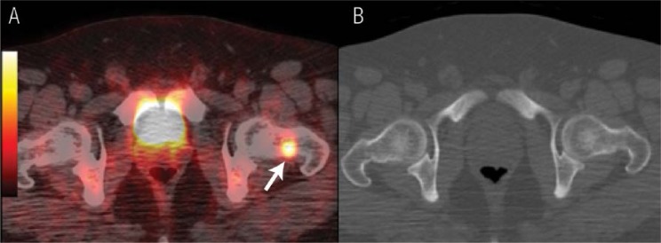
Figure 1 A & B.
18F-fluorodeoxyglucose positron emission tomography (PET) / X-ray computed tomography (CT) for a patient diagnosed with nasopharyngeal carcinoma in which the PET/CT imaging upstaged the disease by revealing a bone lesion that was not detected by CT. A: The fused PET/CT image revealed a focal uptake in the neck of the left femur (arrow). B: The CT image, bone window, did not show any bone abnormalities. The technetium (99mTc)-methylene diphosphonate (MDP) bone scan of this patient was also negative (images not included).

