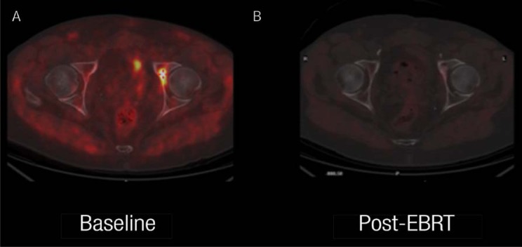
Figure 2 A & B.
18F-fluorocholine positron emission tomography (PET)/X-ray computed tomography (CT) scan in a patient with prostatic adenocarcinoma. This imaging was done to assess the efficacy of therapy in this patient with a single bone metastasis, appearing one year after primary treatment (serum prostate-specific antigen was 2.4 ng/ml). The images are of the fused PET/CT transaxial section at the level of the femoral heads. A: The baseline images showed focal accumulation of the tracer in the left acetabulum, the only site of metastatic disease. B: The favorable response after external beam radiation therapy (EBRT) is shown in this image.

