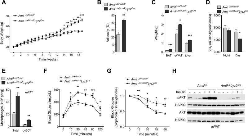
Fig. 5. Myeloid cell-specific deletion of BMAL1 exacerbates metabolic disease.
(A to D) Body weight (A), adiposity (B), tissue weights (C) and oxygen consumption (D) in ArntlLoxP/LoxP and ArntlLoxP/LoxPLyz2Cre mice kept on a 12 hour light-dark cycle fed high fat diet for 19 weeks (n = 4-5 mice per genotype). (E) Total and Ly6Chi macrophage content in eWAT of ArntlLoxP/LoxP and ArntlLoxP/LoxPLyz2Cre mice fed high fat diet (n = 5 mice per genotype). (F and G) Glucose (F) and insulin tolerance (G) tests of ArntlLoxP/LoxP and ArntlLoxP/LoxPLyz2Cre mice fed high fat diet (n = 5-8 mice per genotype). (H) Immunoblots of total and phosphorylated AKT (pAKT) in eWAT of obese ArntlLoxP/LoxP and ArntlLoxP/LoxPLyz2Cre administered intraportal insulin. Representative data of two to four independent experiments (A to C, E to H) are shown as mean ± S.E.M and analyzed using two-tailed Student's t-tests. *P<0.05; **P<0.01; ***P<0.001.

