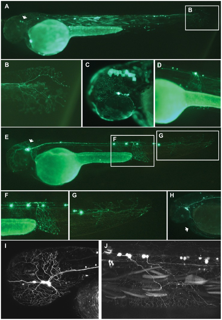
Figure 2. Analysis of Pax6_ciCNE2 and Meis_ciCNE10 in zebrafish embryos.
Pax6_ciCNE2 drives GFP expression in both cranial ganglia (A, C) and spinal cord sensory neurons (B, D) at 48 hrs post fertilization. (B, D) High magnification view of GFP expression detectable in neuronal projections extending into the tail fin and along the spinal cord. (E) Meis_ciCNE10 drives GFP expression in the nervous system at 48 hpf. GFP is strongly expressed in the cell body and in the peripheral process of Rohon-Beard neurons innervating the ventral fin fold (F) and the tail fin (G). (H) GFP expression is detected in trigeminal ganglion neurons and in their process innervating the yolk sac (arrow). (I, J) Confocal analysis showing GFP expression in sensory neurons of the trigeminal ganglion innervating the head as well as in central axons innervating the hindbrain (I) and in projections of Rohon-Beard neurons (J).

